
Pegcetacoplan,
does it really help with Slowed geographic atrophy lesion enlargement in nonexudative age-related macular degeneration?
research showsPegcetacoplan is rated C because it slows geographic atrophy lesion enlargement in nonexudative age-related macular degeneration without established preservation of vision. OAKS and DERBY randomized 637 and 621 participants, respectively. At 24 months, lesion growth was reduced by 22% and 19% with monthly treatment and by 18% and 16% with every-other-month treatment. However, all key visual-function outcomes in both phase 3 trials were null. Slower lesion enlargement is not the same as preserved vision. The current headline claim of slower lesion enlargement warrants C, while claims of preserved vision or visual function warrant D, giving the headline verdict C with 46 points.
ads claimClaims that the drug slows blindness or preserves sight merge imaging lesion growth with actual ability to see. Slower lesion enlargement is not the same as preserved vision, and visual-function endpoints were null in both phase 3 trials. The established headline efficacy is slower lesion enlargement, not vision preservation.
Useful facts when choosing a product
- The United States product Syfovre is given as a 15-mg intravitreal injection into each affected eye once every 25 to 60 days by a qualified physician.
- The treatment does not restore retinal cells or vision already lost. Its demonstrated efficacy is slowing enlargement of geographic atrophy lesion area on fundus imaging.
- New exudative or neovascular AMD can develop during treatment and requires monitoring; anti-VEGF treatment may be needed separately if it occurs.
- Endophthalmitis, retinal detachment, intraocular inflammation, retinal vasculitis or vascular occlusion, and increased intraocular pressure can occur. Sudden pain, redness, photophobia, or vision loss requires urgent ophthalmic evaluation.
Chamgap Semantic Classification Code
Candidate index · review held
M.pegcetacoplan-intravitreal-injection.intravitreal.slowed-geographic-atrophy-lesion-enlargement-in-nonexudative-age-related-macular-degeneration.assess.shamMedicinal interventions > Pegcetacoplan intravitreal injection > Intravitreal > Slowed geographic atrophy lesion enlargement in nonexudative age-related macular degeneration > Association or change assessment > Sham procedure or device
An automated migration candidate, not an issued permanent code; exact claim scope remains under review. This machine-generated migration candidate helps retrieval but is not a permanent assignment. The original verdict ID and URL remain authoritative.
What the research actually shows
OAKS and DERBY were 24-month double-masked phase 3 trials that assigned 1,258 patients with geographic atrophy to intravitreal pegcetacoplan 15 mg monthly, every other month, or sham. OAKS enrolled 637 and DERBY 621. At 12 months, OAKS slowed lesion growth by 21% and 16%, while the 12% and 11% reductions in DERBY were not significant. At 24 months, both trials showed significant imaging effects of 19% to 22% monthly and 16% to 18% every other month. All key visual-function outcomes remained null. New exudative AMD occurred in 11%, 8%, and 2% in the OAKS monthly, every-other-month, and sham groups and in 13%, 6%, and 4% in the corresponding DERBY groups.
Why this is classified as C (46)
Two large phase 3 trials consistently reduced 24-month imaging lesion growth by 16% to 22%, but every key visual-function outcome was null. The current headline lesion-enlargement claim receives C, while vision or visual-function preservation receives D; the imaging ceiling and exudative AMD and endophthalmitis risks give C with 46 points.
Counterpoint. Some patients may value slower imaging progression depending on lesion location, remaining vision, the fellow eye, injection burden, and exudation risk. It should not be described as proven vision preservation, and absolute lesion-area differences and injection risks require discussion with a retinal specialist.
Rejudgment record. Cross-check applied — Rule ①-ⓐ, surrogate only: improvement only in biomarkers that stand in for clinical benefit, such as HbA1c, LDL, blood pressure, intraocular pressure, bone density, laboratory values, or imaging measures, allows a maximum grade of C. Applied C because 24-month geographic atrophy imaging growth decreased while all key visual-function outcomes were null
Sub-claim grades by effect
This ingredient is marketed for several effects. A single overall grade blends strong and weak claims together, so each effect is graded separately here. The overall grade reflects the strongest disconfirming or core claim.
| Effect (sub-claim) | Grade | Basis |
|---|---|---|
| Slowed geographic atrophy lesion enlargement | C | Twenty-four-month imaging lesion growth was reduced by 16% to 22%, but this is an imaging surrogate. Slower lesion enlargement is not the same as preserved vision, and visual-function endpoints were null in both phase 3 trials. |
| Preserved best-corrected visual acuity | D | The key 24-month visual-acuity outcomes in two large phase 3 trials showed no significant benefit. |
| Preserved reading and patient-reported visual function | D | Key visual-function outcomes, including the Functional Reading Independence Index and NEI VFQ-25, were null. |
Cross-check — AI research and Codex final gate
Evidence Table
| Study | Design | Sample | Funding | Endpoint | Result | Weight |
|---|---|---|---|---|---|---|
| Heier JS et al.; OAKS and DERBY study investigators. 2023, OAKS | Twenty-four-month multicenter randomized double-masked sham-controlled phase 3 trial | 637 randomized participants; 614 in the modified intention-to-treat population | Funded by Apellis Pharmaceuticals; multiple company employees were coauthors | Change in fundus-autofluorescence geographic atrophy lesion area and key visual function | At 24 months, lesion growth was reduced by 22% monthly and 18% every other month, but key visual-function outcomes were null. New exudative AMD occurred in 11% and 8% versus 2% with sham. | Confirmatory imaging efficacy with null function |
| Heier JS et al.; OAKS and DERBY study investigators. 2023, DERBY | Twenty-four-month multicenter randomized double-masked sham-controlled phase 3 trial | 621 randomized participants; 597 in the modified intention-to-treat population | Funded by Apellis Pharmaceuticals; multiple company employees were coauthors | Change in fundus-autofluorescence geographic atrophy lesion area and key visual function | The 12-month lesion reduction was not significant, but at 24 months growth was reduced by 19% monthly and 16% every other month. Key visual function was null, and new exudative AMD occurred in 13% and 6% versus 4% with sham. | Delayed replication of imaging efficacy with null function |
| Liao DS et al. 2020, FILLY | Multicenter randomized double-masked sham-controlled phase 2 trial | 246 participants | Funded by Apellis Pharmaceuticals; multiple company employees were coauthors | Twelve-month geographic atrophy lesion growth and visual function | Lesion growth was reduced by 29% monthly and 20% every other month, but vision testing did not improve. | Preceding imaging signal with a functional gap |
Receipt — 3 References
All 3 cited sources were verified for existence at the original page (as of 2026-07-24).
Final verification and publication gate: Codex · Evidence date: 2026-07-24 · Corrections: none
Cite this verdict
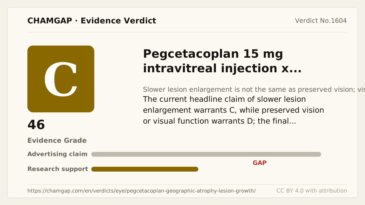
[Chamgap] Pegcetacoplan x slower geographic atrophy lesion enlargement — Evidence Grade C·46. 3 cited sources checked. Source: https://chamgap.com/en/verdicts/eye/pegcetacoplan-geographic-atrophy-lesion-growth/ · CC BY 4.0CC BY 4.0 — free to use with attribution; do not distort grades, numbers, or verdict meaning.
What this document does and does not do
Chamgap is an information source. It reports what research has and has not confirmed; it does not tell readers what to take or buy. That decision belongs to readers and, when needed, medical or legal professionals. This verdict reflects literature available up to the search date and may change as new research appears. Nothing here is medical advice.